
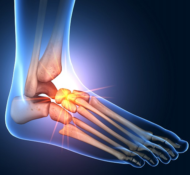
Knee
Anatomy
Conditions / Disorders
Procedures
Anatomy and Joint Of Knee
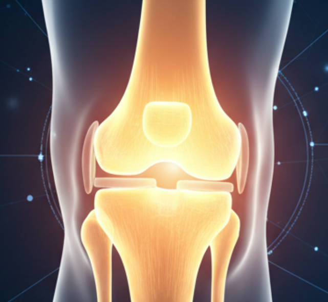
The Knee Joint:
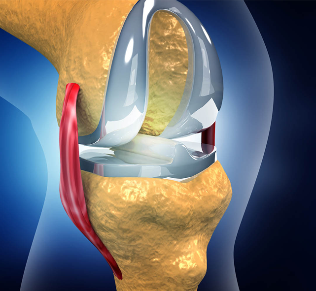
The knee joint is one of the most complex and critical joints in the human body, playing a vital role in movement, weight-bearing, and overall mobility. The knee joint is a hinge-type synovial joint that allows flexion, extension, and slight rotation. It is formed by the articulation of three primary bones:
- Femur (thigh bone): Forms the upper portion of the joint.
- Tibia (shin bone): Forms the lower portion and bears the majority of the body’s weight.
- Patella (kneecap): Protects the joint and enhances leverage during movement.
Key soft tissue structures, including ligaments, cartilage, and tendons, ensure joint stability and smooth movement. The joint is encased in a fibrous capsule lined by synovium, which produces synovial fluid to lubricate the joint.
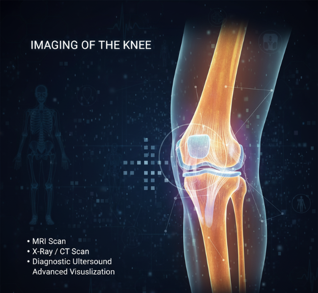
Imaging of the Knee
Medical imaging of the knee joint is essential for accurate diagnosis and treatment planning. It helps healthcare providers determine the cause of knee pain, assess the severity of injuries, and monitor the progress of treatment.
Plain x-rays are a very useful first line investigation, often performed to demonstrate the presence or absence of significant arthritis, but also useful for the identification of loose fragments of bone and other less common conditions such as osteochondritis dissecans and Osgood-Schlatter’s disease.
CT scans offer detailed cross-sectional images of the knee, providing more information about bone injuries and complex fractures. They are particularly useful for preoperative planning and assessing the extent of bone damage.
MRI scanning of the knee, which takes around twenty minutes, produces high quality images of both the bones and the soft tissues within the joint. It is particularly good at imaging the shock absorbing meniscal cartilages and also demonstrates reasonably well the supporting ligaments around the joint, but it is not quite as good at demonstrating injuries to the articular or chondral cartilage.
Ultrasound imaging uses high-frequency sound waves to produce images of soft tissues, such as muscles, tendons, and ligaments. It is useful for diagnosing conditions like tendon tears, ligamentous injuries, and fluid collections within the knee.
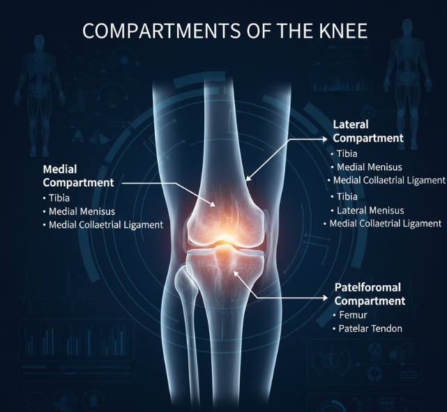
Compartments of the Knee
The knee joint is divided into three main compartments, each playing a unique role in movement and weight-bearing.
Medial Compartment – Located on the inner side of the knee, between the femur and tibia. This compartment bears most of the body’s weight and is the most common site for osteoarthritis.
Lateral Compartment – Found on the outer side of the knee, between the femur and tibia. It helps balance weight distribution and provides stability during side-to-side movements.
Patellofemoral Compartment – Between the patella (kneecap) and the femur. It is essential for knee extension and smooth gliding of the kneecap during activities like climbing stairs or squatting.
Each compartment is lined with articular cartilage to reduce friction and absorb shock. Damage to any of these compartments can cause pain, stiffness, and functional limitations, often guiding treatment strategies such as physiotherapy, injections, or surgical intervention.
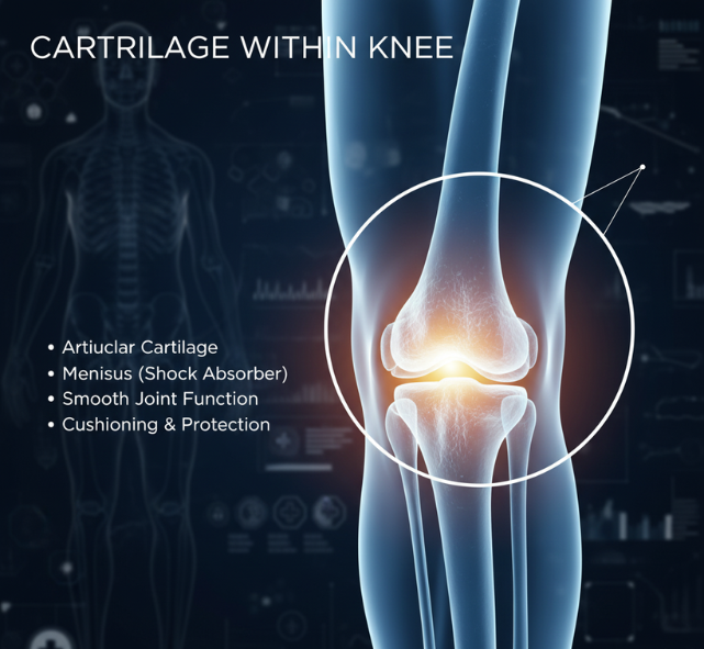
Cartilage within the Knee
The knee contains two types of cartilage: articular cartilage and menisci.
Articular cartilage covers the ends of the femur, tibia, and patella, providing a smooth surface for joint movement. It is a very specialised material, providing lubrication, some shock-absorption, and distributing pressure. It varies in thickness from between a few millimetres to almost a centimetre in depth, and unfortunately is easily damaged. It has limited healing potential and cumulative damage associated with loss of cartilage volume eventually leads to the development of osteoarthritis in old age.
The menisci are C-shaped fibrocartilage structures that act as shock absorbers. They distribute pressure, enhance stability and help the knuckles rotate and slide or glide across each other. They are commonly damaged, particularly in middle age, and this damage is often referred to as torn cartilage or meniscal tear.