


Foot and Ankle
Anatomy
Conditions
Procedures
Anatomy of Foot & Ankle
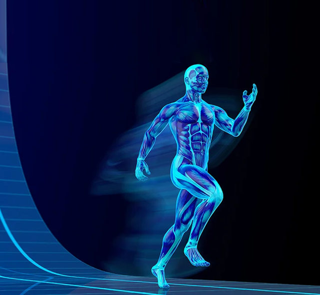
The Ankle & Foot work together to provide mobility, balance, and stability. They allow us to walk, run, and adapt to different surfaces, while supporting the entire weight of the body.
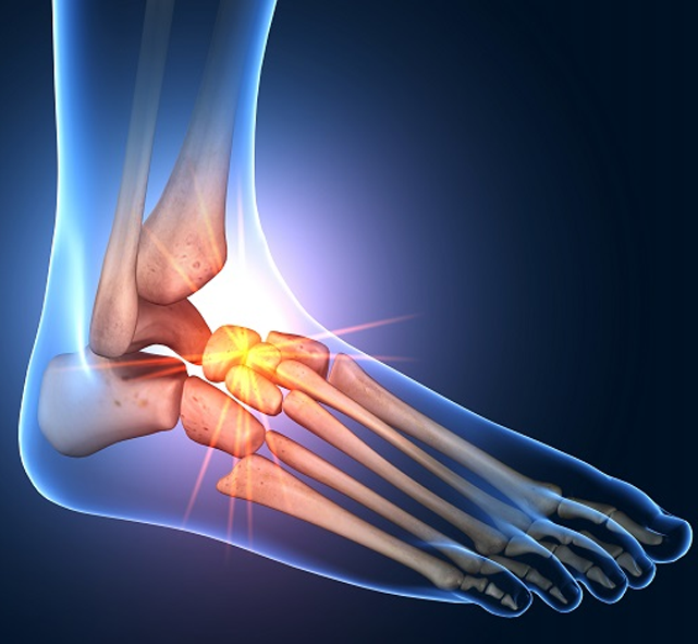
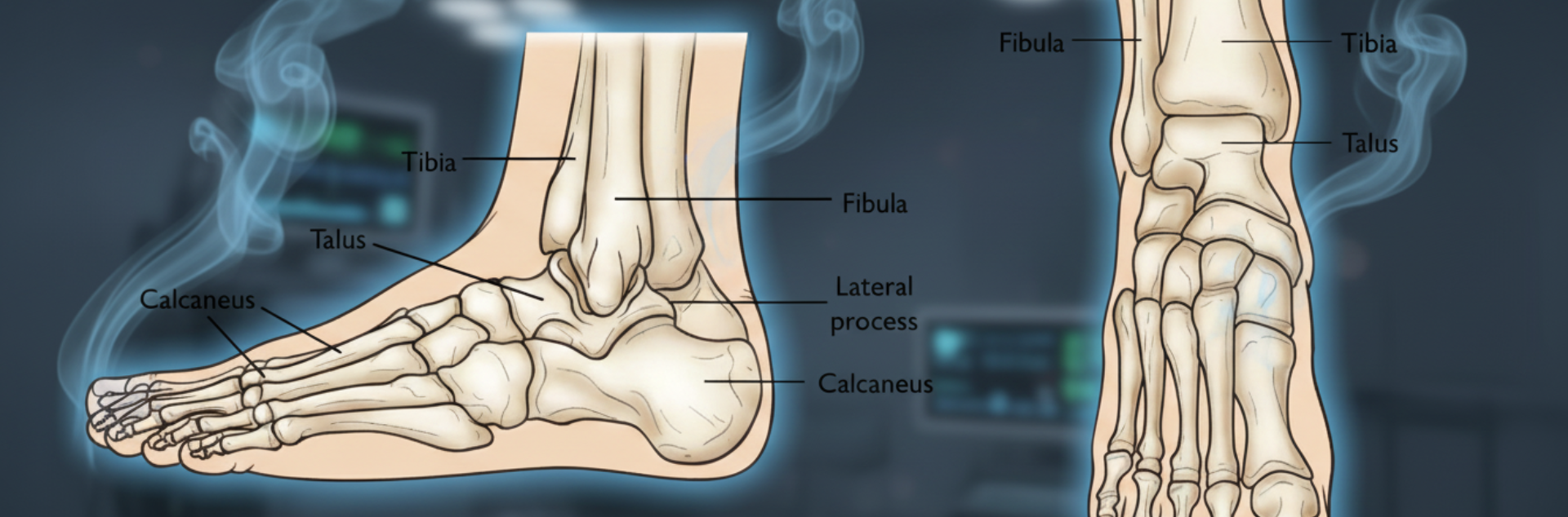
Bones of the Ankle
The ankle joint connects the leg with the foot and is formed by three main bones:
- Tibia (shinbone): Main weight-bearing bone of the lower leg
- Fibula (calf bone): Supports and stabilizes the ankle
- Talus (ankle bone): Sits between the tibia, fibula, and heel bone, allowing up-and-down movement
Three bony prominences around the ankle are important landmarks:
- Medial malleolus– inside of the ankle, formed by the tibia
- Posterior malleolus– back of the ankle, also from the tibia
- Lateral malleolus– outer ankle bone, formed by the fibula
Bones of the Foot
The foot is divided into three regions:
- Hindfoot
- Contains the talus(ankle bone) and calcaneus (heel bone – the largest bone in the foot).
- Midfoot
- Includes the navicular, cuboid, and three cuneiform bones.
- Acts as a bridge between the hindfoot and forefoot.
- Forefoot
- Made up of five metatarsals(long bones forming the arch).
- Phalanges (toes):three bones in each small toe, two in the big toe.
- The big toealso contains two tiny sesamoid bones that help with toe movement.
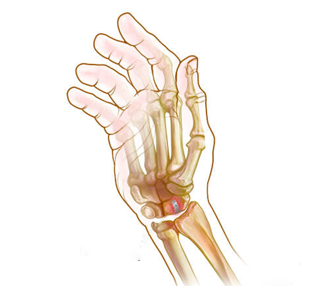
Joints of the Ankle & Foot
There are 33 joints in the ankle and foot, providing a wide range of motion:
- Hinge joints (ankle): Flexion and extension (up and down).
- Gliding joints (hindfoot): Allow sliding movements.
- Condyloid joints (forefoot & toes): Allow bending, side-to-side, and spreading of toes.
All joint surfaces are covered with articular cartilage, a smooth, flexible tissue that cushions and reduces friction. The joints are further lubricated by synovial fluid for easy movement.
Soft Tissues of the Ankle & Foot
- Cartilage: Smooth tissue covering bone ends, acting as shock absorbers.
- Ligaments: Strong bands connecting bones to bones, providing stability.
- Plantar fascia– runs from heel to forefoot, supports the arch.
- Lateral ligaments– outside of the foot, stabilize ankle movement.
- Medial ligaments– inside of the foot, allow controlled motion.
- Muscles: About 20 muscles help with movement and support.
- Anterior tibial muscle: Up-and-down movement of the foot.
- Posterior tibial muscle: Supports the arch.
- Peroneal muscles: Control movement on the outer ankle.
- Extensors: Lift the toes during walking.
- Flexors: Stabilize toes against the ground.
- Tendons: Connect muscles to bones.
- Achilles tendon: Strongest tendon, attaches calf muscle to heel bone.
- Other key tendons: Peroneals, tibialis anterior and posterior.
- Bursae: Small fluid-filled sacs that reduce friction between tendons, bones, and skin.