

The elbow joint is a complex hinge joint that connects the upper arm to the forearm. It allows two primary types of movement:
- Flexion and extension (bending and straightening the arm)
- Pronation and supination (rotation of the forearm turning the palm up or down)
The elbow is highly stable due to its strong bony architecture, ligaments, and surrounding muscles.
Bones of the Elbow
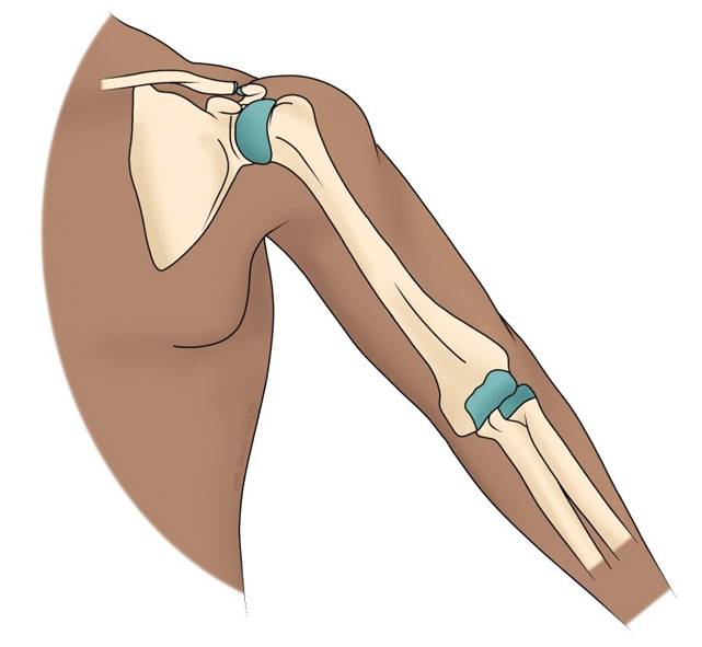
The elbow joint is formed by the articulation of three bones:
- Humerus – the upper arm bone.
- Ulna – the inner and larger bone of the forearm, lying on the little-finger side.
- Radius – the outer bone of the forearm, lying on the thumb side.
Together, they form three joints enclosed in a single capsule:
- Humeroulnar joint – hinge joint between trochlea of humerus and trochlear notch of ulna (main flexion/extension).
- Humeroradial joint – between capitulum of humerus and head of radius.
- Proximal radioulnar joint – pivot joint between radial head and radial notch of ulna (allows pronation/supination).

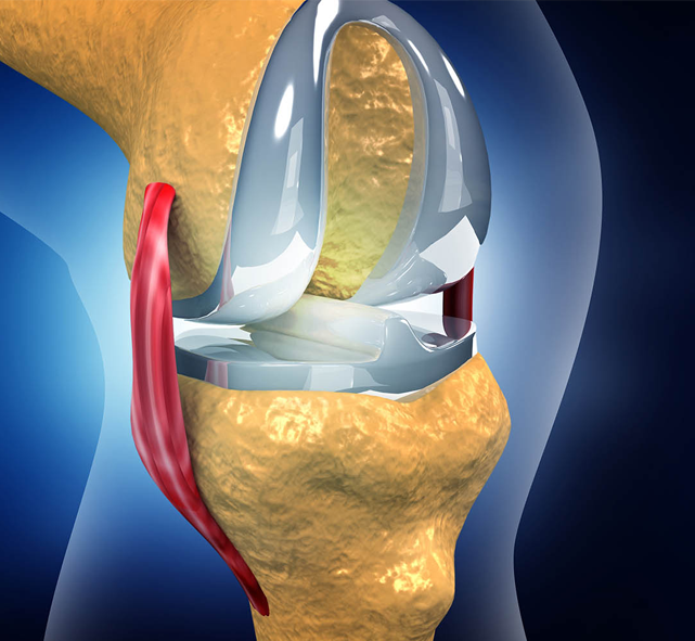
Soft Tissues of the Elbow
- Articular cartilage: covers the ends of the bones, reducing friction and absorbing stress.
- Joint capsule: encloses all three articulations in a single synovial capsule, providing protection and lubrication.
Ligaments of the Elbow
The elbow is reinforced by several strong ligaments:
- Ulnar Collateral Ligament (Medial Collateral Ligament) – runs from humerus to ulna, stabilizes the inner side of the joint.
- Radial Collateral Ligament (Lateral Collateral Ligament) – runs from humerus to radius, stabilizes the outer side.
- Annular Ligament – encircles the head of the radius and holds it in the radial notch of the ulna, allowing rotation.
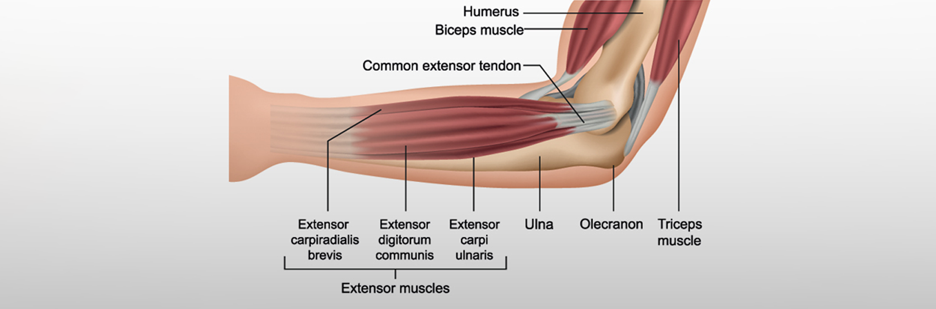
Muscles of the Elbow
Muscles around the elbow provide movement and stability:
- Flexors (bend the elbow): primarily biceps brachii, brachialis, and brachioradialis.
- Extensors (straighten the elbow): primarily triceps brachii and anconeus.
- Pronators (rotate palm downward): pronator teres and pronator quadratus.
- Supinators (rotate palm upward): supinator and biceps brachii.

Tendons of the Elbow
- Biceps tendon – attaches biceps muscle to the radius, allowing supination and flexion.
- Triceps tendon – attaches triceps muscle to the ulna, responsible for extension.
- Common flexor tendon – origin of wrist/finger flexors from the medial epicondyle of humerus.
- Common extensor tendon – origin of wrist/finger extensors from the lateral epicondyle of humerus.
Nerves of the Elbow
Important nerves passing around the elbow include:
- Median nerve – passes through the cubital fossa, supplies forearm flexors.
- Ulnar nerve – runs behind the medial epicondyle (“funny bone”), supplies intrinsic hand muscles.
- Radial nerve – runs around the lateral elbow, supplies extensors of forearm.
Blood Vessels of the Elbow
The elbow has a rich vascular supply:
- Brachial artery – main artery of the arm, bifurcates into radial and ulnar arteries at the level of the elbow.
- Radial and Ulnar arteries – supply the forearm and hand.
- Venous drainage – via deep veins (brachial, radial, ulnar) and superficial veins (cephalic, basilic, median cubital vein).