
Shoulder Anatomy
The shoulder is the most flexible joint in the human body, enabling a wide range of movements such as forward flexion, abduction, adduction, external rotation, internal rotation, and full 360° circumduction.
Because of this exceptional mobility, the shoulder joint is also considered the most insecure joint in the body. However, stability is provided through the combined support of ligaments, muscles, tendons, and the joint capsule.
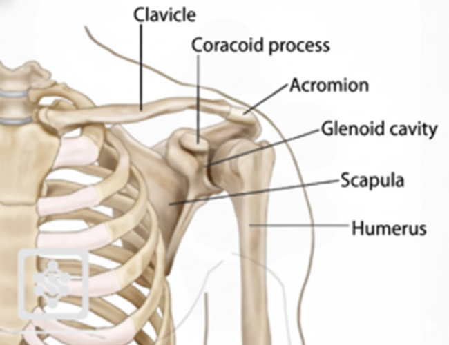
Bones of the Shoulder
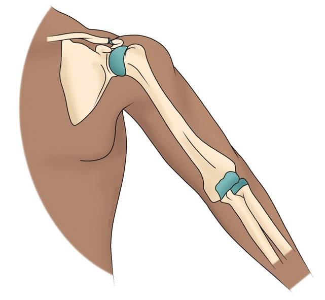
The shoulder is a ball-and-socket joint formed by three main bones:
- Humerus – the upper arm bone, whose rounded head forms the “ball.”
- Scapula – the triangular shoulder blade, with a shallow depression called the glenoid cavity that forms the “socket.”
- Clavicle – the collarbone, which connects the shoulder to the sternum.
Together, these structures form key joints:
- Glenohumeral joint – main ball-and-socket articulation between humeral head and glenoid cavity.
- Acromioclavicular joint – between clavicle and acromion process of scapula.
- Sternoclavicular joint – between clavicle and sternum.
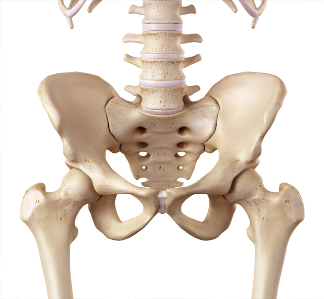
Key structures include:
- Femoral head and acetabulum
- Articular cartilage
- Ligaments (iliofemoral, pubofemoral, ischiofemoral)
- Labrum
- Surrounding muscles such as gluteals, iliopsoas, and quadriceps
Soft Tissues of the Shoulder
- Articular cartilage: smooth tissue covering bone ends to reduce friction and absorb shock.
- Glenoid labrum: a ring of fibrous cartilage that deepens the glenoid cavity and enhances joint stability.
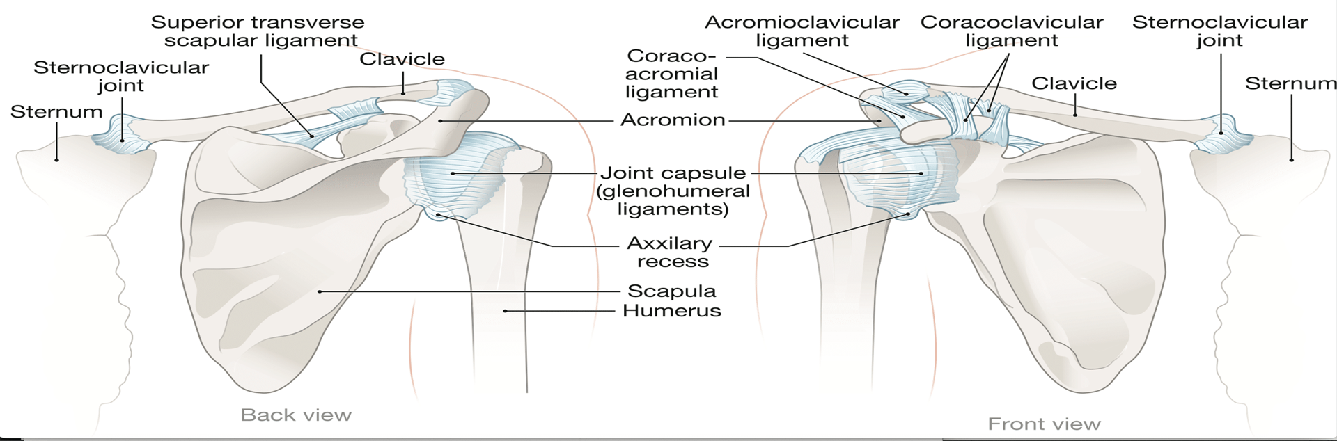
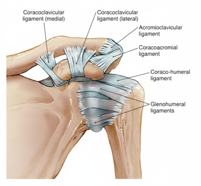
Ligaments of the Shoulder
Shoulder ligaments connect bone to bone, forming a stabilizing capsule:
- Coracoclavicular ligaments – clavicle to coracoid process.
- Acromioclavicular ligament – clavicle to acromion process.
- Coracoacromial ligament – connects acromion and coracoid process.
- Glenohumeral ligaments (3) – form a capsule around the joint, preventing dislocation and maintaining stability.
Muscles of the Shoulder
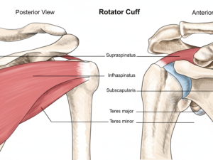
The rotator cuff is the key muscle group providing stability and movement. It consists of four muscles:
- Supraspinatus
- Infraspinatus
- Teres minor
- Subscapularis
Together, they stabilize the humeral head in the glenoid cavity while enabling mobility.
The deltoid muscle, the largest and strongest muscle of the shoulder, provides power for lifting and overhead movements.
Tendons of the Shoulder
- Rotator cuff tendons – connect cuff muscles to the humerus, crucial for joint stability.
- Biceps tendons – connect the biceps muscle to the shoulder (long head and short head).
Nerves of the Shoulder
The brachial plexus (a bundle of nerves from the neck) passes through the shoulder to supply the arm. Key nerves include:
- Musculocutaneous
- Axillary
- Radial
- Ulnar
- Median
These nerves transmit motor commands and sensory feedback.
Blood Vessels of the Shoulder
Blood supply is delivered mainly by the subclavian artery, which becomes the axillary artery in the armpit and the brachial artery further down the arm.
Main veins include:
- Axillary vein → drains into subclavian vein
- Cephalic vein → drains into axillary vein
- Basilic vein → drains into axillary vein